Tag Archives: Doctor of Philosophy

Endothelial Progenitor Cells For Injured Tissue Repair

Endothelial Progenitor Cells Role on Injured Tissue

Endothelial Progenitor cellsThe endothelial progenitor cells are sourced from the bone marrow and have been found to have the ability to proliferate and differentiate in mature endothelial cells. However, these cells are not only sourced from the bone marrow alone but can also be found in large proportions in non-marrow sources like spleen which particularly has been found to be rich in EPCs. Isolated spleen-derived mononuclear cells, pre-selected with an endothelial cell medium, demonstrated endothelial cell characteristics and formed tubular-like structures. There are hopes that these cells can be used to sufficiently improve re-endothelialization and lessen neointima formation after carotid artery injury. In a trial, intravenous transfusion of spleen-derived EPCs in splenectomized mice showed special homing to the injured area. However, these results were only achieved when the host organ was removed. Thereafter, it was suggested that removal of the spleen prolonged the EPC time in circulation, which may result in a change of surface markers on the cell because of homing signals of the injury site thus favoring recruitment to the ischemic area rather than preferential homing to the organ of origin. With researchers still working to establish the mechanism with which the EPCs repair damaged tissues; there is hope that these cells can be useful in treatment of injured tissues.

Apart from the crucial role of maintaining the cardiovascular homeostasis that the vascular endothelial cells play, they also provide a physical barrier between the vessel wall and lumen. The endothelium also secretes a number of mediators that regulate platelet aggregation, coagulation, fibrinolysis, and vascular tone. However crucial functions the endothelium plays, it may lose its physiological properties and hence termed endothelial dysfunction. Incase this occurs it will not be able to promote vasodilation, fibrinolysis, and anti-aggregation as it normally does to ensure sound vascular health. Endothelial cells secrete several mediators that can alternatively mediate either vasoconstriction, such as endothelin-1 and thromboxane A2, or vasodilation, such as nitric oxide (NO), prostacyclin, and endothelium-derived hyperpolarizing factor. Nitric oxide is the chief contributor to endothelium-dependent relaxation in conduit arteries; however the contribution of endothelium-derived hyperpolarizing factor predominates in smaller resistance vessels.

Restores endothelial functions

The endothelial progenitor cells are essential as they are immature but with the ability of differentiating into mature endothelial cells and hence may help restore the endothelial functions in case of injuries that may result in endothelial dysfunction. The release of growth factors and cytokines may cause vascular injury and tissue ischemia which will in turn mobilize endothelial progenitor Cells which will specifically home in on the ischemic sites to stimulate compensatory angiogenesis once in the peripheral circulation.

Furthermore, endothelial progenitor cells forms part of a pool of cells able to form a cellular patch at sites of endothelial injury, thus working directly to achieve the homeostasis and repair of the endothelial layer. Endothelial progenitor cells have now been identified to be playing a major role in cardiovascular biology, as a matter of fact, the extent of the circulating EPC pool is now considered a mirror of cardiovascular health. Practically all risk factors for atherosclerosis have been linked to declining population of circulating endothelial progenitor cells or their dysfunction. The increase in population of the circulating endothelial progenitor cells have been linked to decreased cardiovascular mortality.

Endothelial progenitor cellsOne of the common diseases of the cardiovascular is atherosclerosis which is characterized by leucocyte infiltration, smooth muscle cell accumulation, and neointima formation. Basically atherosclerosis is a cardiovascular inflammatory disease. It has been shown that the activation and damage of the endothelial layer is what causes the development of lesions. Recent studies have disputed an earlier notion that the adjacent intact endothelium replaces the damaged endothelial cells. These studies have demonstrated the recruitment and incorporation of vascular progenitor cells into atherosclerotic lesions and thus providing evidence in support of the role of vascular cells in the development of the disease. The incorporation of endothelial progenitor cells into mice showed promising results. In a model of transplant atherosclerosis, regenerated endothelial cells from arterial grafts were found to originate from recipient circulating blood but not the remaining endothelial cells of the donor vessels. It was also found that the endothelial monolayer in a vein graft three days after surgery was completely lost and later replaced by circulating endothelial progenitors.

The endothelial progenitor cells are able to mediate vascular repair and attenuate the progression of this disease even when there is a continued vascular injury. treatment of chronic injuries have been done with endothelial progenitor cells in mice in trials , however the mechanism involved is still a mystery but it is clear that these EPCs contribute a big deal to the restoration of the injured endothelial layer. In one example, intravenous infusion of spleen-derived mononuclear cells increases endothelium-dependent vasodilatation in atherosclerotic mice, signifying that progenitor cells play an important role in repairing the vascular injury.

Finally, for more information about bone marrow transplant and stem cell transplantation, visit www.awaremednetwork.com. Dr. Dalal Akoury has been practicing integrative medicine for years; she will be able to help. You can also visit http://www.integrativeaddiction2015.com and learn more about the upcoming Integrative Addiction Conference 2015. The conference will deliver unique approaches to telling symptoms of addiction and how to assist patients of addiction.

Endothelial Progenitor Cells Role on Injured Tissue


Facebooktwitterpinterestlinkedin

Exploring Spinal Cord Injuries

The Evolving Science Of Spinal Cord Injuries

Spinal cord InjuryThe most important structure that plays the role of a link between the body and the brain is the spinal cord. It extends from the medulla oblongata of the brain through to the level of the first lumbar vertebrae. It is a cylindrical structure of nervous tissue that is composed of white and grey matter. On each of its sides, two consecutive rows of nerve roots emerge. These roots distally join forming the 31 pairs of spinal nerves. It is housed within the vertebral column. The spinal cord makes up approximately only 2% of the central nervous system but has very vital functions.

A spinal cord injury refers to any damage to any part of the spinal cord or damage of nerves at the spinal canal’s end. Below the site of the injury, there often occur permanent changes in sensation, strength and other body functions.

The subject of spinal cord injuries is not a new one. Many people are familiar with it and understand its financial implications. Many families have had to put up with hefty hospital bills after a spinal cord injury of a member. Special equipment like wheelchairs have had to be purchased to assist victims of spinal cord injury. Some victims even lose their jobs after a spinal cord injury due to inability to perform their tasks at the work place. This is a very serious financial blow to them considering the fact that they require large amounts of money to lead a normal life.

There is a complete relationship between the physical, financial, emotional and social implications of spinal cord injuries. For example when one suffers a spinal cord injury, they get paralyzed which calls for the purchase of special equipment. In some cases paralysis may lead to loss of a job. When one loses a job, they become emotionally affected and thus their social life is also greatly affected.

Spinal cord injuries are of different levels and seriousness depending on the site of injury. The vertebrae that make up the spinal column are grouped into sections. Since spinal injuries affect body functions below the site of injury, the higher the sight of injury, the more severe the dysfunction that results.

Spinal cord injury cure has been the focus of science for so long. New strategies are being developed and old strategies are being revised to make them better and improve their efficacy.

Regenerative strategies

The last two decades have witnessed tremendous efforts in attempt to enhance regeneration of spinal cord axon though many techniques. These techniques include neutralization of neurite inhibition, synthetic channel implantation, various cell transplantation and administration of neurotrophic factors. Some of these strategies have been applied to animal models and they have been so promising to the extent that their potential human application is being explored by clinicians. The main factor limiting recovery from a spinal cord injury has been attributed to the failure of the central nervous system to regenerate. All these strategies aim at making it possible for the axons in the central nervous system to regenerate. Expectations become high with the identification of growth inhibitory molecules in the central nervous system. It was thought that neutralizing these factors would allow for functional axonal regeneration in the central nervous system. But as happens to most seemingly bright researches, the dark side finally showed in this. There exist mixed results of therapeutic approaches that were based on this assumption. Neurons are suggested to differ in their regenerative abilities through similar extracellular environments by recent data. These neurons have also been shown by recent data to undergo a developmental loss of intrinsic regenerative ability. Intrinsic regenerative abilities are mediated by factors that include expression of:

  • Cytoskeletal proteins mediating the axon growth mechanics
  • Receptors for inhibitory molecules
  • Molecules in the intracellular signaling cascades mediating response to chemoattractive and chemorepulsive cues.
  • Surface molecules permitting adhesion of axon to cells in the growth path

Sharply contrasting to axon development, its regeneration involves internal protrusive forces. Micro tubules generate these forces either by transporting other skeletal elements like neurofilaments to the tip of the axon or through their own elongation. It is the complexity of the regeneration program that casts a dark shadow on the progress.

Multi- cell therapy

Spinal cord injuryTransplantation of cells replaces the damaged neural tissues and restores function after spinal cord injury. Successful results have been obtained with different cell types including adult neural stem cells, mesenchymal stem cells, fetal tissue, embryonic stem cells and myelin producing cells.

In transplants of fetal tissue combined with neurotrophic factors, axonal growth has been seen. Transplanting polymer guiding channels with Schwann’s cells also helps in achieving novel axonal growth towards a cell transplant.

It is important to note that enough immune suppression is needed lest the cell transplant will be rejected. Attention is currently focused on mesenchymal stem cells to try and circumvent this immunological rejection of transplanted cells. After transplantation, they differentiate into desired cells.

Even though multiple studies have shown success of these multi-cell strategies, it is still not clearly understood what mechanisms lead to functional improvement following transplant.

For more information about bone marrow transplant and stem cell transplantation, visit www.awaremednetwork.com. Dr. Dalal Akoury has years of experience in integrative medicine and will be of assistance.

While at it, visit http://www.integrativeaddiction2015.com to learn about the upcoming integrative addiction conference 2015. The conference will deliver unique approaches to telling symptoms of addiction and how to assist patients of addiction.

The Evolving Science Of Spinal Cord Injuries


Facebooktwitterpinterestlinkedin

Power of Light Therapy

The Healing Power of Light Therapy

Light TherapyThe benefits of light are many but to most people exposure to sunlight has only one benefit associated with vitamin D. however there is much worse views that have been propagated in relation to exposure to sunlight and while the fact that the UV rays exposure may cause skin cancer and harmful sunburn, the cosmetic industry has blown this issue out of proportion and the reality however is the intensity of the UV rays that causes such harmful effects is rarely achieved. The benefits of exposure to light have been overlooked perhaps clouded by the negative comments that had been propagated in the past. Exposure to light has many benefits.

Ultraviolet light as a medical treatment has been used since the beginning of the century. The technique is merely using UV light to stimulate the immune system and various enzyme systems. It is a tested and proven therapy that has accomplished incredibly miraculous cures with absolutely no side effects, and yet until recently it has been suppressed and ignored by American medicine.

UV light for disinfection

The use of UV light in infection isn’t new; it has been used for several years and is still being used for the same purpose. The approach to use of UV light in disinfection requires use of little sophisticated equipment and works by stimulating the body’s own immune response and various enzyme systems. When this UV light is used for disinfection, a small amount of blood is drawn from the body and is treated with UV light (photoluminescence) and then the blood is reinjected into the bloodstream. When this is done the body’s defenses are rapidly organized to destroy all invading organisms whether viral, bacterial or fungal. According to William C. Douglass, M.D, an author of a book entitled; Into the Light, a husband and wife, who both had a flu one treatment completely, reversed the system in both patients within two hours. The author however states that in case of serious infections, marked reduction in toxic symptoms is observed in 12 to 48 hours.

Another doctor, Dr. Jonathan V. Wright, MD believes that the benefits of the UV light far more outweigh its risks. He believes that UV rays from the sun are the best source of the vitamin D your body needs to ward off cancer and dozens of other health problems. But that’s just the beginning of what ultraviolet light can do. Not only is it an extremely effective disinfectant with the ability to kill bacteria, viruses, and fungi in the air and on surfaces but UV light also has the potential to prevent and even cure infections and diseases that other treatments are powerless against.

to many people the use of UV light in medical treatment may sound as a new technology but the reality however is that this treatment has been used since 19th century in prevention and treatment of diseases and hence isn’t a fresh area of research. The first experiment on the medical applicability of the UV light was done on the patients with lupus and sepsis. A breakthrough in the use of UV light saw a Danish physician named Niels Ryberg Finsen win a Nobel Prize for his work with UV light and the treatment of the disease in 1903.

Now that the benefits of light is known to you, there is a concern, why do we need light therapy when we can get all the benefits from the sun source UV light?

To answer this, I won’t dispute that the sun sourced UV light is the best but there are places or seasons when the sun isn’t seen and therefore there is need for a light therapy to help people in times of these times to evade dangers of deficiency of light. This is where light therapy comes in.

Light therapy also known as bright light therapy involves the use of light boxes emitting full-spectrum light similar in composition to sunlight. Daily exposure to this bright light is the treatment method most often recommended for patients with Seasonal Affective Disorder (SAD). SAD is a form of depression that occurs as a result of reduced exposure to sunlight in the fall, winter and spring. When the SAD symptoms become more severe doctors recommend the light therapy to help reverse the condition.

Light TherapyNormally, during the treatment the patient will sit in front of a light box for thirty minutes a day during the season when light is inadequate. The treatment works best when the patient goes for it immediately after waking up. The patient can read, write or eat when sited before the light but it is advisable for the patient to stop looking directly at the light box. Light therapy is believed to work by its effect on brain chemicals that play a role in regulating mood. This treatment can relieve symptoms within a few days, but sometimes takes as long as two weeks or more.

Conditions that can be treated with light therapy

The light therapy can be used for the following disorders; skin disorder psoriasis and some other skin conditions, as well as for a type of cancer called cutaneous T-cell lymphoma. This light therapy is also effective in treatment of depression, insomnia, anxiety and can also be used to counter the effects of antidepressants.

Eating disorders like bulimia have been also treated by light therapy. However this treatment is still being studied for treatment of obsessive-compulsive disorders, premenstrual syndrome, jet lag, Parkinson’s disease, dementia and attention deficit hyperactivity disorder (ADHD).

Finally, Dr. Dalal Akoury (MD) is an experienced doctor who has been in the frontline in pursuit of integrative medicine that works best. He runs a website that equips readers of better ways to overcome not only drug addiction but also serious health problems that have caused nightmares to the world population. Get in touch with her today and learn more.

The Healing Power of Light Therapy


Facebooktwitterpinterestlinkedin

Understanding the History of Gotu Kola

Understanding the History of Gotu Kola: The Magic of Gotu Kola

Understanding the History of Gotu Kola

Understanding the History of Gotu Kola as it was used then and the today’s application

Gotu kola is a kind of vegetation which has been instrumental in the traditional treatment of various diseases in most countries including India, China, and Indonesia. Scientifically it is known as Centella asiatica. Some of the major areas of applications included healing of wounds, improvement of mental clarity as well as treating skin conditions such as leprosy and psoriasis. This plant has been singled out to one of the most used herbs in providing alterative treatment for so many health complications. There is a lot being said about this herb and we want to get the clear understanding of the history of gotu kola as it was then and how it is beneficial currently. The roles of this herb are endless. Some people use it to treat respiratory infections such as colds and in the past China used it primarily for that among many other ways. And because of its ability to offer treatment solutions, it was nick named “the fountain of life”. The motivation of this nick name was because legend has it that an ancient Chinese herbalist lived for more than two centuries (200 years) and his long life was credited to gotu kola.

Archaeologically, gotu kola has also been used to treat some STI including syphilis, hepatitis, stomach ulcers, mental fatigue, epilepsy, diarrhea, fever, and asthma. Currently this herb is penetrating in to the American and Europe market where it is being used to treat varicose veins and chronic venous insufficiency. This is a health condition where blood pools in the legs. Besides that it’s also used in ointments to treat psoriasis and help heal minor wounds thereby keeping the good health of the skin. Experts at AWAREmed Health and Wellness Resource Center under Doctor Akoury’s care are reiterating that gotu kola should not be mistaken for kola nut (Cola nitida) because they are not the same. Doctor Akoury explains that unlike kola nut, gotu kola does not have caffeine and is not a stimulant.

Understanding the History of Gotu Kola: Plant Description

  • Gotu kola is a perpetual plant native to India, Japan, China, Indonesia, South Africa, Sri Lanka, and the South Pacific.
  • It is member of the parsley family it has no taste or smell.
  • It thrives in and around water.
  • It has small fan shaped green leaves with white or light purple-to-pink flowers, and small oval fruit.
  • The leaves and stems of the gotu kola plant are used as medicine.

Understanding the History of Gotu Kola: Medicinal Uses and Treatment Indications

Gotu kola has very good medicinal characteristics and can be very useful in treatment of so many health complications including the following:

Venous insufficiency and varicose veins – When blood vessels lose their elasticity, blood pools in the legs and fluid leaks out of the blood vessels. That causes the legs to swell (venous insufficiency). Several small studies suggest gotu kola may help reduce swelling and improve blood flow. In a study of 94 people with venous insufficiency, those who took gotu kola saw their symptoms improve compared to those who took placebo. In another study of people with varicose veins, ultrasound tests showed that people who took gotu kola has less leakage of fluid.

Wound healing and skin lesions – Gotu kola has chemicals called triterpenoids. In animal and lab studies, these compounds seem to help heal wounds. For example, some studies suggest that triterpenoids strengthen the skin, boost antioxidants in wounds, and increase blood supply to the area. Based on these findings, gotu kola has been applied to the skin, or used topically, for minor burns, psoriasis, preventing scars after surgery, and preventing or reducing stretch marks.

Anxiety – These same chemicals triterpenoids seem to decrease anxiety and increase mental function in mice. One human study found that people who took gotu kola were less likely to be startled by a new noise than those who took placebo. Since the “startle noise” response can be a way to tell if someone is anxious, researchers think that gotu kola might help reduce anxiety symptoms. However the dose applied for this study was very high making it impossible to say how gotu kola might be used to treat anxiety.

Scleroderma – A single study of 13 women with scleroderma found that gotu kola decreased joint pain and skin hardening, and improved finger movement.

Insomnia – Gotu kola acts as a sedative when given to animals in tests. Because of that, it is sometimes suggested to help people with insomnia. But no human studies have been done to see whether it works and whether it’s safe.

Understanding the History of Gotu Kola: Dosage and Administration

Gotu kola is available in teas and as dried herbs, tinctures, capsules, tablets, and ointments. Products should be stored in a cool, dry place and used before the expiration date on the label. At no given time should gotu kola be used for patients under the age of 18 years old. Nonetheless as for the adults, the standard dose of gotu kola (Centella asiatica) will fluctuate depending on what kind you use and what you use it for. Getting your doctor’s opinion would be highly recommended in choosing the right dose for you.

Understanding the History of Gotu Kola: Side Effects

Side effects are rare but may include skin allergy and burning sensations with external use, headache, stomach upset, nausea, dizziness, and extreme drowsiness. These tend to happen with high doses of gotu kola.

Pediatric use – Gotu kola is not recommended for children.

Geriatric Use – The dosage of 65 years old and above should take a lower dose of gotu kola. Your health care provider can help you determine the right dose for you, which can be increased slowly over time.

Understanding the History of Gotu Kola: Interactions and Depletions

Gotu kola may interact with the following medications:

Drugs that affect the liver – Gotu kola contain elements which are likely to injure the liver of the patient. It must therefore be noted that taking it along with some other medications that also can harm the liver may cause liver damage.

Cholesterol-lowering drugs (including statins) – In some studies where animal were involved it was established that gotu kola raised cholesterol levels of those animals. Therefore it can may also do the same (raise cholesterol levels) in humans. However this is still a hypothesis since it is yet to be confirmed.

Diabetes medications – While carrying out a study in animal it was established that, gotu kola seems to increase blood sugar levels. Individuals with diabetes should not take gotu kola without first talking to their doctor.

Diuretics (water pills) – Gotu kola seems to act like a diuretic, meaning it helps the body get of excess fluid. Taking diuretic medications and gotu kola could cause your body to lose too much fluid, upsetting the balance of electrolytes you need. The same is true of taking gotu kola with herbs that have diuretic effects, such as green tea, astragalus or gingko.

Sedatives – Because gotu kola acts like a sedative, it might make some drugs taken for anxiety or insomnia stronger. The same is true for herbs taken for anxiety or insomnia, such as valerian.

Understanding the History of Gotu Kola: The Magic of Gotu Kola


Facebooktwitterpinterestlinkedin

Human Adipose-Derived Mesenchymal Stem Cells Reduce Inflammation

Human Adipose-Derived Mesenchymal Stem Cells Reduce Inflammation modulates Regulatory T Cells in Rheumatoid Arthritis

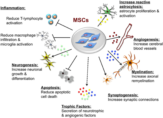
Mesenchymal stem cells are a rare type of cells that are multi-potent and can be isolated from the adipose tissues, bone marrow among other crucial sources.  These cells have very unique properties that have seen them become targets of very many medical therapies especially in treating degenerative diseases like aging and even joint diseases like arthritis. These cells have been found to be also very effective in modulating the functions of immune cells, including T cells, B cells, natural killer cells, monocyte or macrophages, dendritic cells, and neutrophils. T cells, activated to perform a range of different effector functions, are the primary mediators of many autoimmune and inflammatory diseases as well as of transplant rejection and graft-versus-host disease. Some of the well-defined T-Cell effector cells include; CD4+ (T helper cell) subsets Th1, Th2, and Th17 cells and cytotoxic T lymphocytes which are derived from antigen-specific activation of simple CD8+ precursors. In addition, naturally occurring and induced regulatory T cells represent CD4+ and CD8+ T-cell phenotypes that potently suppress effector T cells to prevent autoimmunity, maintain self-tolerance, and limit inflammatory tissue injury. The ability of adipose-derived mesenchymal stem cells to suppress the effector T-Cells and limit inflammation has made them very effective in treating rheumatoid arthritis which is characterized with inflammations around the joints. Typically, many immune-mediated diseases entail an imbalance between regulatory T cells and effector T cells of one or more phenotypes. Mesenchymal Stem Cells broadly suppress T-cell activation and proliferation in a controlled environment outside of a living organism via (Vitro) a plethora of soluble and cell contact-dependent mediators.  These mediators may work in two ways; first they can work directly on T cells or indirectly via modulation of antigen-presenting cells and other accessory cells. Mesenchymal Stem Cells administration has also been shown to be variably associated with beneficial effects in autoimmune and transplant models as well as in several human clinical trials. However, in a small number of studies Mesenchymal Stem Cells administration has been found to heighten T cell-mediated tissue injury. The multiple effects of Mesenchymal Stem Cells on cellular immunity may reflect their diverse influences on the different T-cell effector subpopulations and their capacity to specifically protect or induce regulatory T-cell populations.

Mesenchymal Stem Cells

 Mesenchymal stem cell modulation of T cell-mediated immune responses

To repeat for emphasis, the T cells are the primary cellular effectors of the adaptive immune system and their functional properties are central to antigen specificity and memory associated with cognate immunity, this therefore means that to a greater extent they are responsible for rejection of implanted cells of varied specificity.  Antigen-specific activation and differentiation of naïve T cells result in the generation of a range of T-cell phenotypes that may be defined by the acquisition of characteristic cytokine secretion profiles, cytolytic mechanisms, or counter-regulatory properties.

Following antigen-specific adaptive immune responses, a small proportion of activated T cells persist as memory cells and have the capacity to respond more rapidly and potently to secondary encounters with the same antigen. These memory cells may retain the effector phenotype imprinted upon them during primary activation. When these memory cells are appropriately coordinated and regulated, the diversity of T-cell effector phenotypes allows immune protection against a multitude of pathogenic microorganisms while maintaining self-tolerance and homeostasis. On the other hand, over exuberant pro-inflammatory T-cell responses may lead to auto-immune and allergic diseases, including multiple sclerosis, inflammatory bowel disease, type 1 diabetes mellitus, and asthma. Therefore it is crucial to have a modulatory strategy on the effector T-cells. Furthermore, life-saving treatments such as allogeneic bone marrow and solid organ transplantation may be complicated by alloantigen-specific T-cell immune responses, resulting in graft-versus-host disease (GvHD) or transplant rejection.

Study finding

Mesenchymal Stem Cells and InflammationIn a research study done at School of Medicine, University of Seville, Seville, Spain by Gonzalez-Rey E, Gonzalez MA, Varela N, O’Valle F, Hernandez-Cortes P, Rico L, Büscher D,  and Delgado M  it was found that human adipose-derived mesenchymal stem cells suppressed the antigen-specific response of T cells from patients with rheumatoid arthritis. Human adipose-derived mesenchymal stem cells inhibited the proliferative response and the production of inflammatory cytokines by collagen-activated CD4 and CD8 T cells. On the contrary, the numbers of IL10-producing T cells and monocytes were significantly augmented upon human adipose stem cell treatment. The suppressive activity of human adipose-derived mesenchymal stem cells was cell-to-cell contact dependent and independent. Human adipose-derived mesenchymal stem cells also stimulated the generation of FoxP3 protein-expressing CD4 (+) CD25 (+) regulatory T cells, with the capacity to suppress collagen-specific T cell responses. Lastly, human adipose-derived mesenchymal stem cells down regulated the inflammatory response and the production of matrix-degrading enzymes by synovial cells isolated from patients with rheumatoid arthritis.

The study had sought to find out the immunosuppressive activity of human adipose-derived mesenchymal stem cells on collagen-reactive T cells from patients with rheumatoid arthritis.  The method used involved investigating the effects of human adipose-derived mesenchymal stem cells on collagen-reactive RA human T cell proliferation and cytokine production as well as effects on the production of inflammatory mediators by monocytes and fibroblast-like synoviocytes from patients with rheumatoid arthritis. Stem cell therapy is a promising approach to treatment of degenerative diseases like arthritis but still you will need an expert in degenerative medicine. Dr. Dalal Akoury (MD) an expert in integrative and regenerative medicine will be able to help. Visit us at AWAREmed Health and Wellness Resource Centre at Myrtle Beach, South Carolina

Human Adipose-Derived Mesenchymal Stem Cells Reduce Inflammation modulates Regulatory T Cells in Rheumatoid Arthritis

Facebooktwitterpinterestlinkedin